Nerves are wiring our body. How much do we know about the nerve microstructure?
Improve models + accuracy of diffusion MRI: we need to know the true 3D microstructure of the BRAIN. Does diabetes influence the radius, trajectory and organization of myelinated axons in human peripheral NERVES?
Description /program:
Part 1: Diffusion magnetic resonance imaging (MRI) is a non-invasive imaging modality very suitable for imaging brain, Diffusion MRI allows us to extract statistics about axon microstructure in the different parts of the brain. With diffusion MRI, we can assess the manifestation of neurological diseases and monitor the changes in brain microstructure. This leads to improved diagnosis and treatment of neurological diseases.
Diffusion MRI relies on accurate models for brain microstructure. Currently used models are based on measures obtained from histology and electron microscopy, which are high-resolution modalities, but unsuitable for 3D inference. Those models are therefore not accounting for 3D effects such as bending and diameter variation. To improve our models, and the accuracy of diffusion MRI, we need to know the true 3D microstructure of the brain.
BREAK with fruit and beverage
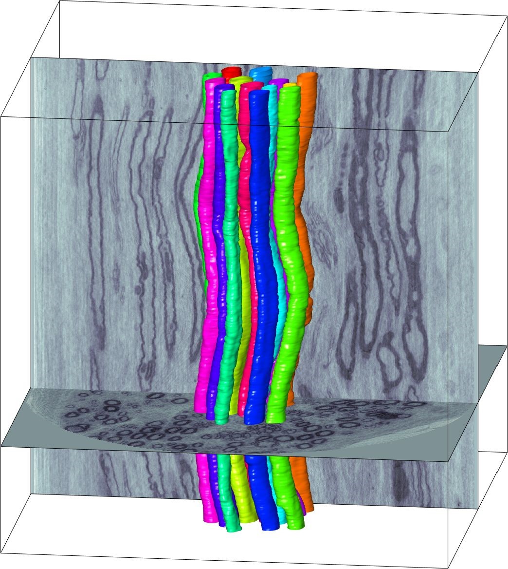
Part 2: Advances in tomography allow us to visualize the 3D microstructure of nerve tissue. We are often interested in whether a disease affects the tissue and changes its microstructure. Such a hypothesized correlation is exemplified in the following questions: Does diabetes influence the radius, trajectory and organization of myelinated axons in human peripheral nerves? How does multiple sclerosis influence the microstructure of the white matter?
In these questions, the hypothesized change in the fibre microstructure is subtle and can´t be revealed by visual inspection, or by conducting measurements in only a few places. Therefore, automated methods leading to the quantification of fibre geometry are required for establishing the correlation between the fibre microstructure and the disease.
Speakers:
Associate professor at DTU Compute Tim B. Dyrby timd@drcmr.dk
Associate professor at DTU Compute Vedrana Andersen Dahl vand@dtu.dk
Organizer: Finn Gustafsson, IDA IT, figu@dtu.dk
Gratis for medlemmer af IDA IT og DANSK IT
This event is closed for registration.
Information